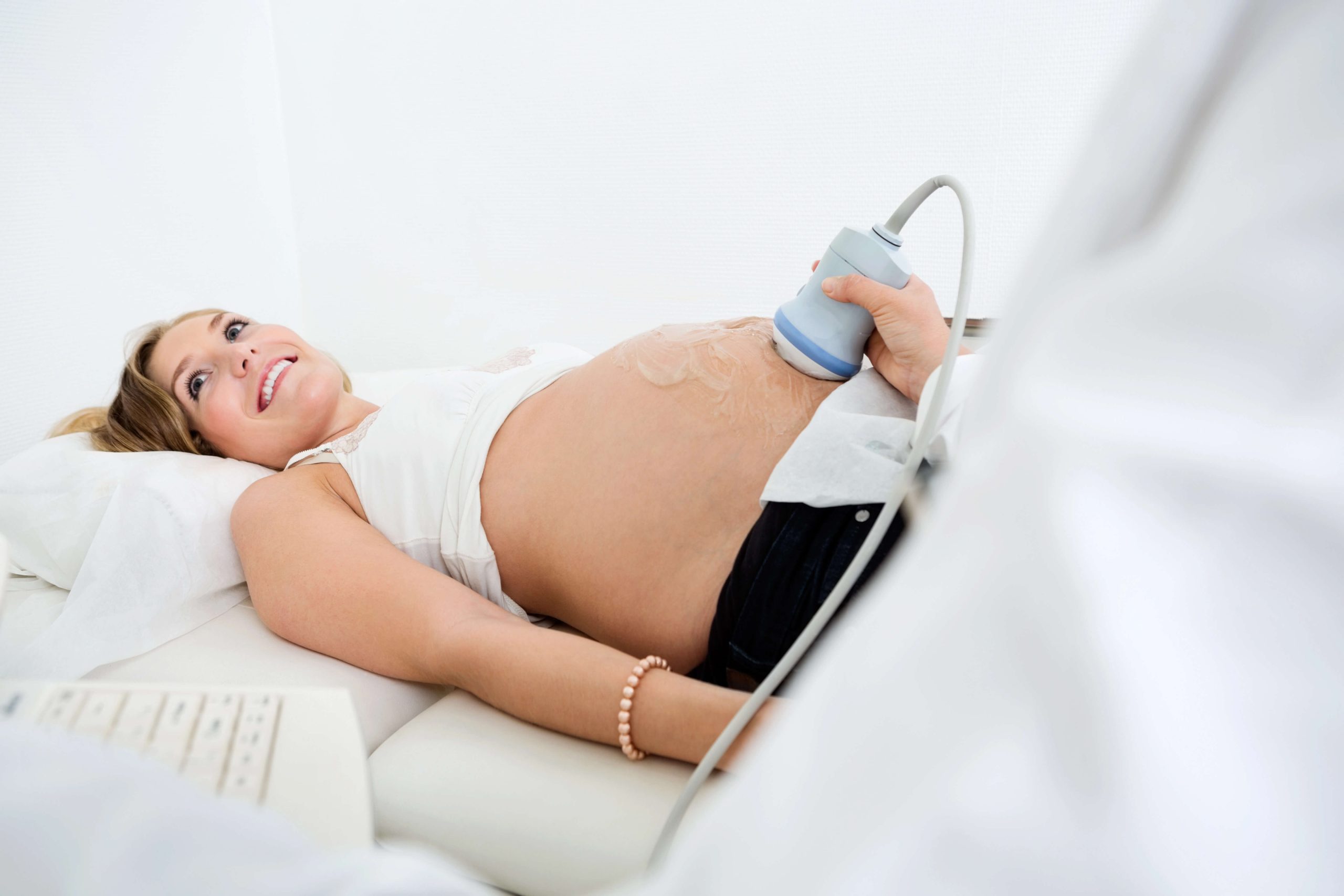
Pregnancy is a journey filled with anticipation, excitement, and a fair share of anxiety. For expectant parents, one of the most reassuring aspects of this journey is seeing their baby grow and develop through ultrasound scans.
The wellbeing is also known as Growth scan is often conducted around 26 weeks of pregnancy up to term. Usually, wellbeing scans were suggested at 28, 32 and 36 weeks of pregnancy. There are few reasons where wellbeing scan is ordered by the Obstetrician such as antepartum bleeding, reduced fetal movements, preterm rupture of the membranes and suspected small baby based on physical examination. In high risk pregnancy example when the mother is having preeclampsia which resulting in small baby, there will be few interval scans suggested to monitor the growth as well as the blood flow.
For certain twin pregnancy such as identical twin or Monochorionic diamniotic twin (MCDA Twin), the follow up will be more compares to singleton pregnancy because in some cases, about 10-15% of these twin pregnancies will develop twin-to-twin transfusion syndrome (TTTS). This happened because both babies are sharing the same placenta resulting one of the babies receive lesser blood flow. It is crucial for this type of pregnancies to be monitored more closely to check for early signs of TTTS or other complications for better outcomes.
Wellbeing scan involves assessing the placental position and fetal presentation, measuring fetal size, identifying any abnormalities, evaluating the amniotic fluid level, and assessing the blood flow in brain and umbilical cord.
Placental position
Assessment of placental location is crucial in third trimester if the patient is diagnosed with a low-lying placenta or placenta previa during the anomaly scan. There will be few subsequent scans to ensure the placenta is not covering the internal os. The best approach to assess the internal os in third trimester is by transvaginal scan as posterior placenta is difficult to assess from transabdominal approach. Maternal obesity and presence of uterine fibroids are another reason why transvaginal scan is preferred.
Fetal presentation
The optimal fetal position for vaginal delivery is when the baby is head down, facing your back and the back of its head ready to enter the pelvis. This is referred to as cephalic or occiput anterior presentation, and most babies settle into this position by the 36th week of pregnancy. Breech presentation is one of the risk factors for developmental dysplasia of the hip (DDH).
Fetal growth
To ensure the baby is growing at health rate, we can estimate the baby weight by measuring the head circumference (HC), abdominal circumference (AC) and femur length (FL). At late pregnancy, HC measurement will be more difficult to obtain as the fetal head is low in maternal pelvis.
Fetal abnormalities
Fetal abnormalities, which typically become apparent or develop after the second-trimester scan, often affect the genitourinary tract, central nervous system (CNS), and heart. For instance, fetal cardiac rhabdomyoma which, although rare, yet it is the most common cardiac tumor found in fetuses. To take note that Wellbeing scan is not an Anomaly scan. Due to fetal size, limited space, and the bone are more ossified causing more prominent shadowing, some structures are difficult to assess especially in late trimester.
Amniotic fluid level
Checking the amount of amniotic fluid surrounding the baby, which is crucial for their lung development. It can be done by measuring the amniotic fluid index (AFI). Low amniotic fluid or oligohydramnios can be related to pathology in fetal urinary system and premature rupture of membrane. Polyhydramnios is defined as AFI value is more than 25 cm. Polyhydramnios may associate with maternal diabetes, congenital malformations of gastrointestinal tract such as duodenal atresia, fetal infections, and chromosomal and genetic abnormalities.
Middle cerebral artery (MCA) and Umbilical artery (UA) Doppler assessment
Umbilical artery Doppler assessment is crucial to endure that baby is receiving enough blood and nutrients from the mother. In fetuses with intrauterine growth restriction (IUGR), the blood flow waveform could be abnormal. The middle cerebral artery (MCA) Doppler is important in assessing fetal anemia or hypoxia. One of the common causes of fetal anemia is Rhesus (Rh) incompatibility. Rhesus incompatibility is you have rhesus negative blood (RhD negative) and your baby has rhesus positive blood (RhD positive).
The Wellbeing Scan offers numerous benefits to both the parents and the healthcare providers. One of the primary benefits is the peace of mind it provides to expectant parents by seeing a healthy, active baby can significantly reduce anxiety. Early identification of potential problems allows for timely intervention, which can improve outcomes for both the mother and the baby.
When it comes to selecting a provider for your Wellbeing Scan, choosing a reputable clinic with experienced sonographers is key. At our clinic, we offer high-quality pregnancy scans, with a dedicated team focused on ensuring each scan is a positive and reassuring experience for expectant parents. We understand the emotional journey of pregnancy and are committed to delivering the highest standard of care.
If you’re expecting a baby and considering a Wellbeing Scan, we invite you to contact our Dublin Ultrasound Clinic. Our team here in Dundrum is ready to support you every step of the way, providing the information and reassurance you need to ensure a healthy, stress-free pregnancy.

